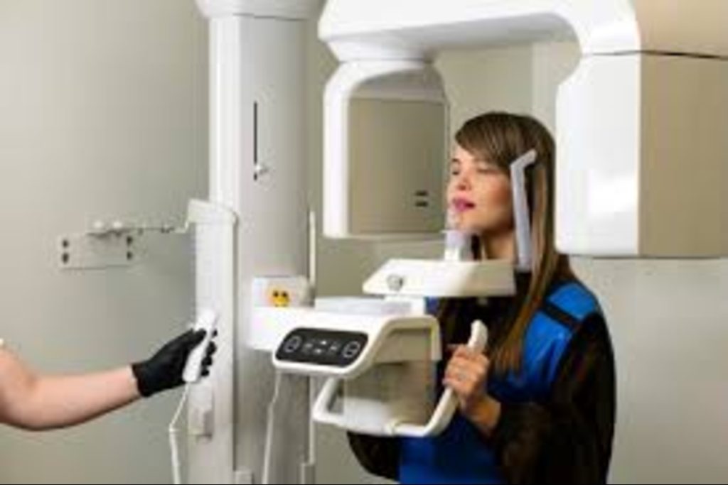
A regular dental X-ray shows teeth and surrounding structures in two dimensions. Cone beam computed tomography (CBCT) creates a three-dimensional view that can reveal details hidden by overlapping structures.
A dentist in Coquitlam may recommend CBCT when conventional images do not provide enough detail to answer a specific question about tooth position, bone, or nearby structures. The scan isn’t needed for every concern; it provides information that could affect diagnosis or treatment planning.
What Does CBCT Show That a Regular X-Ray May Not?
Traditional dental X-rays compress anatomy into a flat image. CBCT lets the clinician view teeth, bone, and nearby structures from multiple angles. It can clarify root position, bone dimensions, or a tooth’s relationship to a nerve or sinus.
When comparing CBCT vs. dental X-ray, the right approach depends mainly on the information needed during the assessment. Standard X-rays often suffice for routine exams, cavities, and common dental concerns. Consider CBCT when a 2D image leaves an important question unanswered, and the result could change care.
The three-dimensional view can reduce uncertainty in selected cases, but it may also reveal incidental findings that need further review. Its value lies in helping answer a specific clinical question, not simply providing more detail.
When Might a Dentist Recommend a Scan?
Dentists may consider three-dimensional imaging for unusual root anatomy, suspected jawbone changes, certain injuries, or procedures near important structures. Recommend it when the additional information could support diagnosis or treatment planning.
For impacted wisdom teeth, CBCT may clarify the roots’ relationship to nearby nerves when regular X-rays leave uncertainty. This is one application of cone beam CT for wisdom tooth imaging, though it isn’t necessary for every wisdom tooth assessment.
How Is CBCT Used for Dental Implants?
Before implant placement, the clinician needs to assess jawbone dimensions and nearby structures. CBCT for implant planning provides this information in three dimensions and helps identify anatomical limits, especially when bone grafting or a more complex approach is considered.
The examination, existing images, and site complexity determine whether CBCT is warranted. Imaging supports planning but does not replace clinical judgment or guarantee a particular outcome.
What About Radiation Exposure?
CBCT uses ionizing radiation, generally more than a single dental X-ray. Exposure varies by machine, settings, and the area being scanned. Concerns about radiation from 3D dental scans are valid, particularly for children or people who have undergone repeated imaging.
The scan should be justified by a clear clinical need, with the field of view limited to the area required. Mention any possibility of pregnancy and relevant previous imaging.
Existing images may help avoid an unnecessary repeat scan. For children and teens, the decision requires particular care because developing tissues are more sensitive to radiation.
What Happens Before and After the Scan?
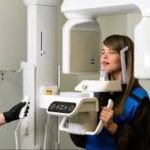
The clinician should explain the scan’s purpose and how the findings could affect treatment. You may need to remove glasses, jewelry, or hair clips. During imaging, the machine rotates around your head while you remain still. Your dentist then reviews the images alongside your dental history and examination.
A cone beam CT scan in Coquitlam may be appropriate when you need three-dimensional information for diagnosis or planning. Coquitlam Dental Group lists Cone Beam CT Imaging among its technologies, but whether a scan is suitable depends on your individual clinical needs.
What Should You Discuss With Your Dentist?
If you are discussing CBCT with a dentist near you, a few questions can help clarify why the scan has been recommended:
- What information is the scan expected to provide?
- Could a regular dental X-ray provide enough information?
- Which part of the mouth or jaw needs to be included?
- How could the findings affect diagnosis or treatment?
Understanding these points can help you make an informed decision about your care.
Clarify Your Imaging Options
A 3D scan is worthwhile when it provides information that can guide a clinical decision. Schedule a consultation with Coquitlam Dental Group to discuss your concerns and imaging options. Get clear answers about what is needed for your care.
FAQs
How is CBCT different from a regular dental X-ray?
A dental X-ray creates a two-dimensional image, while CBCT produces a three-dimensional view of roots, bone, and nearby structures. It helps in selected cases, but it isn’t necessary for every dental problem.
Does a 3D dental scan use radiation?
Yes. Exposure varies by scan type, settings, and field of view. Use CBCT when the expected benefit justifies the exposure, and limit the scanned area to what is needed.
Is CBCT always needed before dental implants?
No. The implant site, anatomy, examination, and existing images determine whether CBCT could improve planning. It is not automatically required for every implant.
Can a regular X-ray be enough to assess wisdom teeth?
Sometimes. Conventional images often provide enough information for assessment and planning. Consider CBCT when the tooth’s position or relationship to nearby structures remains unclear, and the findings could change treatment.